
The Spirotome
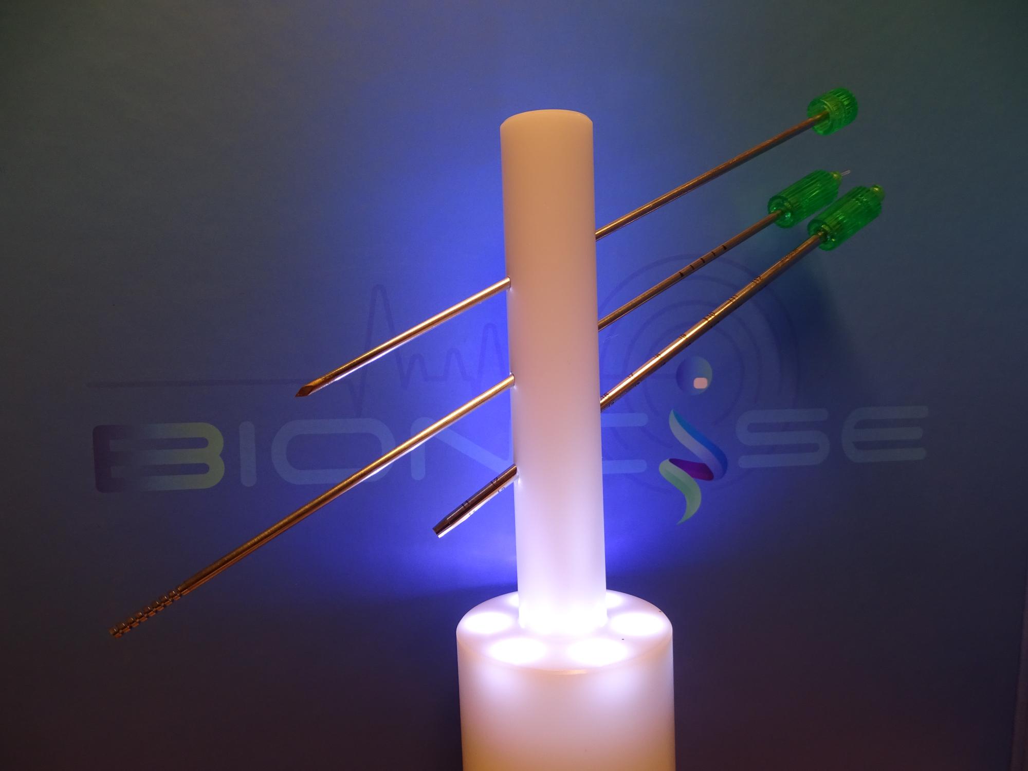
The Spirotome is the main product to acquire high quality soft tissues for engineering and liquid phase analysis. The ready-to-use kit contains a trocar, receiving needle with helix, cutting cannula, and release element. The kit is offered steam sterilized for single use applications.
In general, the mounted trocar and cutting cannula are positioned in front of the target. The trocar is replaced by the receiving needle with helix. The helix is turned clock-wise to enter the target up to the distance needed with a maximum penetration of 20 mm. Then the sample is cut from the surroundings by turning the cutting cannula clockwise over the helix until the distal ends meet. The receiving needle can now be retracted. The sample is removed by the release element. After that, the cutting cannula can be removed or used as starting point for the next biopsy.
The Bioncise products comprise the Spirotome and Corticore families. They contain variations of the base products for various clinical applications.
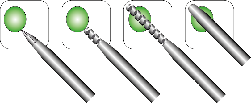
4 simple steps to operate the Spirotome:
1. Position the trocar with cutting cannula up to the target.
2. Replace the trocar with the receiving needle.
3. Turn the helix into the target (maximally 20 mm)
4. Cut the sample by turning the cutting cannula.

Contents of the kit:
Look how it works: